
To that end, we designed a new rotational support plate (RSP) (Fig. Although there are many surgical approaches and internal fixation methods for this type of fracture, methods that have the advantages of simple surgical approaches lead to small intraoperative trauma and have reliable internal fixation effects have not been reported. Although fibula osteotomy provides more exposure of the posterolateral plateau, fracture healing at the osteotomy site and the stability of the lateral structure of the knee joint are not ideal. The fracture needs to be fixed through the supra fibula space, and the fixation effect is often not ideal. In a relatively simple anatomical structure, the anterolateral approach can expose only part of the lateral plateau due to the obstruction of the fibula, so it is difficult to expose the fracture.
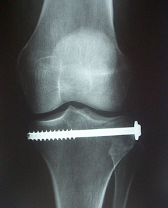
:max_bytes(150000):strip_icc()/tibialplateau-56a6d9223df78cf7729089cd.jpg)

Because there are important neurovascular branches in the surgical area (such as the common peroneal nerve), careful anatomical separation is required. With the traditional treatment method, the posterior or posterolateral approach is usually used in the knee joint to directly separate and expose the posterolateral platform, reposition the structures under direct vision and place the internal fixator behind the tibia for fixation. If good reduction and fixation are not observed after a fracture, significant discomfort and dysfunction occur. The posterolateral tibial plateau is essential for stabilizing the posterolateral knee joint, especially when the knee joint is flexed. These fractures are often combined with fractures of other parts of the tibial plateau, accounting for 14.3–44.2% of all tibial plateau fractures. In tibial plateau fractures, the posterolateral segment of the tibia plateau is frequently affected and challenging to treat. With our newly designed RSP and special pressurizer, posterolateral tibial plateau fractures can be easily and effectively reduced and fixed through the anterolateral approach, which serves as a novel treatment for posterolateral tibial plateau fractures.
CLOSED FRACTURE TIBIAL PLATEAU SKIN
Skin necrosis, incision infections, and fixation failure did not occur during the follow-up period. The average KOOS QOL score was 88 (range, 69–93). The average KOOS sport/recreation score was 83 (range, 70–90). The average KOOS ADL score was 91 (range, 74–97). The average KOOS Pain score was 91 (range, 72–97). The average KOOS Symptoms score was 90 (range, 75–96). The average HSS score was 91 (range, 64–98). At the last follow-up, the average knee range of motion was 138° (range, 107–145°).

The average bony union time was 3.2 months (range, 3–4.5 months). The average follow-up time of all patients was 16.5 months (range, 12–25 months). The Hospital for Special Surgery (HSS) knee score and Knee Injury and Osteoarthritis Outcome Score (KOOS) were used to evaluate knee function at the last follow-up. Postoperative rehabilitation was advised, knee X-rays were taken at follow-ups, and fracture healing, complications, and knee range of motion were assessed. Methodsįrom May 2016 to January 2019, the data of 12 patients with posterolateral tibial plateau fractures treated with the RSP and special pressurizer in our hospital were retrospectively analyzed. This study details the technique of treating these fractures with the RSP and special pressurizer and provides the outcomes. This method is advantageous because it leads to little trauma, involves a simple operation, and has a reliable fixation effect. We designed a new rotational support plate (RSP) and a special pressurizer that can fix the fracture directly via the anterolateral approach. Although there are many surgical approaches and fixation methods for the treatment of these fractures, all of these methods have limitations.


 0 kommentar(er)
